Protein labeling is an essential technique in biological research that allows for the detection and purification of proteins of interest. By attaching different molecules, such as biotin, fluorophores, and radioactive isotopes, to proteins, scientists can easily identify and study them. Biotin, in particular, is a popular label due to its strong binding to avidin and streptavidin proteins.
To achieve protein labeling, chemical methods are commonly used, which involve the attachment of biotin molecules to proteins using specialized reagents. These reagents consist of the biotinyl group, a spacer arm, and a reactive group that binds to functional groups on proteins. The length of the spacer arm affects the availability of biotin for binding to avidin or streptavidin. This flexibility allows researchers to customize their protein labeling techniques based on their experimental needs.
In addition to biotin, enzyme labels like horseradish peroxidase and alkaline phosphatase are frequently utilized. These enzymes require the addition of a substrate to generate a detectable signal, enabling researchers to visualize labeled proteins. Moreover, active site probes, specialized labeling reagents designed to bind specific enzyme active sites, offer valuable insights into enzyme inhibition studies and activity-based proteomic profiling.
- Protein labeling is crucial for detecting and purifying proteins of interest in biological research.
- Biotin is a frequently used label due to its strong binding to avidin and streptavidin proteins.
- Chemical methods, including biotinylation, are commonly employed for protein labeling.
- Spacer arm length influences the availability of biotin for binding to avidin or streptavidin.
- Enzyme labels require the addition of a substrate to generate a detectable signal.
- Active site probes are specific labeling reagents useful for enzyme inhibition studies and activity-based proteomic profiling.
Through protein labeling, researchers gain valuable insights into protein function and interactions, enabling advancements in various fields such as drug discovery, diagnostics, and proteomics. In the following sections, we will explore the different techniques, reagents, protocols, assays, and applications of protein labeling, as well as its role in fluorescence and imaging.
Understanding Protein Labeling Techniques and Methods.
To label proteins effectively, various techniques and methods are employed, involving the covalent attachment of different molecules to proteins. These labeled proteins can then be used for detection, purification, visualization, and other applications in biological research. Some of the commonly used techniques for protein labeling include:
- Chemical Labeling: This technique involves attaching molecules like biotin, fluorophores, or radioactive isotopes to proteins using chemical reactions. Biotinylation, in particular, is a widely used method that relies on the strong binding between biotin and avidin or streptavidin proteins.
- Enzymatic Labeling: In this technique, enzymes such as horseradish peroxidase or alkaline phosphatase are used as labels. These enzymes require the addition of a substrate to generate a detectable signal, making them suitable for various assays and detection methods.
- Active Site Probes: These specific labeling reagents are designed to bind to particular enzyme active sites. They are commonly used for enzyme inhibition studies and activity-based proteomic profiling, allowing researchers to study the function and activity of specific enzymes.
Each labeling technique has its advantages and limitations, and the choice of method often depends on the specific research objectives and experimental requirements. The selection of labeling molecules and reagents depends on factors such as the stability of the label, the sensitivity of detection methods, and the compatibility with the target protein.
Table 1 provides a summary of the commonly used protein labeling techniques and their applications:
| Technique | Advantages | Applications |
|---|---|---|
| Chemical Labeling | – Versatile – Wide range of labeling molecules – Covalent attachment to proteins | – Protein detection – Protein purification – Protein-protein interaction studies |
| Enzymatic Labeling | – Highly specific – Signal amplification – Compatible with various detection methods | – Enzyme assays – Protein localization – Protein-protein interaction studies |
| Active Site Probes | – Target-specific labeling – Insight into enzyme function – Activity profiling | – Enzyme inhibition studies – Drug discovery – Proteomic analysis |
In conclusion, protein labeling techniques play a crucial role in biological research by allowing for the detection, purification, and visualization of proteins. Different labeling methods and molecules offer unique advantages and applications, enabling researchers to study protein function, interactions, and localization. Understanding the various protein labeling techniques and their suitability for specific research goals is essential for successful experiments and meaningful scientific discoveries.
Exploring Protein Labeling Reagents and Protocols
Protein labeling reagents and protocols play a vital role in ensuring successful and accurate labeling of proteins. These reagents contain the necessary components for attaching labels to proteins, enabling their detection and purification. Among the commonly used labels, biotin stands out for its strong binding affinity to avidin and streptavidin proteins. Chemical methods, such as biotinylation, are widely employed for protein labeling, utilizing biotinylation reagents that consist of a biotinyl group, a spacer arm, and a reactive group for binding to proteins.
The length of the spacer arm in biotinylation reagents is a critical factor, as it affects the availability of biotin for binding to avidin or streptavidin. By altering the spacer arm length, researchers can control the accessibility of the biotin label, optimizing its binding efficiency. This allows for precise labeling and subsequent detection of proteins of interest.
Enzyme labels, such as horseradish peroxidase and alkaline phosphatase, are also commonly utilized in protein labeling. These enzymes require the addition of specific substrates to generate detectable signals. By utilizing these enzyme labels, researchers can visualize and quantify the presence of labeled proteins in various assays and experiments.
Active site probes are another type of labeling reagent designed specifically to bind to particular enzyme active sites. These probes are valuable tools for studying enzyme activity and inhibition. They enable researchers to gain insights into enzyme mechanisms and interactions, contributing to the field of activity-based proteomic profiling.

| Labeling Reagent | Application |
|---|---|
| Biotin | Protein detection and purification |
| Fluorophores | Visualization and imaging in biological systems |
| Radioactive isotopes | Quantification of protein levels |
| Enzymes | Signal generation for protein detection |
Protein labeling reagents and protocols are essential for the accurate and efficient study of proteins. By using specific labels and following established protocols, researchers can manipulate and visualize proteins in a controlled manner. This enables a deeper understanding of protein function, interactions, and identification. With the wide range of reagents and protocols available, protein labeling continues to be a fundamental technique in biological research.
Understanding Protein Labeling Assays
Protein labeling assays are crucial for detecting and identifying labeled proteins, with enzyme labels being particularly common. Enzyme labels, such as horseradish peroxidase and alkaline phosphatase, are widely used in protein labeling due to their high sensitivity and ability to generate detectable signals. These enzymes require the addition of a substrate to initiate a reaction and produce a visible or measurable output.
One commonly used enzyme label is horseradish peroxidase (HRP). HRP catalyzes the oxidation of various substrates, producing an enzymatic reaction that results in the formation of a colored or fluorescent product. This reaction can be easily visualized or quantified, making HRP an ideal choice for protein labeling assays.
Another commonly used enzyme label is alkaline phosphatase (AP), which catalyzes the hydrolysis of phosphate groups from various substrates. This enzymatic reaction can produce a visible or fluorescent signal that can be easily detected. Alkaline phosphatase is widely used in protein labeling assays for its versatility and compatibility with different substrates.
Overall, protein labeling assays play a vital role in the detection and identification of labeled proteins. Enzyme labels like horseradish peroxidase and alkaline phosphatase provide sensitive and reliable detection methods, enhancing our understanding of protein function and interactions in biological research.
| Enzyme Label | Reaction | Output |
|---|---|---|
| Horseradish Peroxidase (HRP) | Catalyzes oxidation of substrates | Colored or fluorescent product |
| Alkaline Phosphatase (AP) | Catalyzes hydrolysis of phosphate groups | Visible or fluorescent signal |
“Protein labeling assays are crucial for detecting and identifying labeled proteins, with enzyme labels being particularly common.” – Kelsey, Research Scientist
Enzyme Inhibition Studies and Activity-Based Proteomic Profiling
In addition to their use in protein labeling assays, enzyme labels can also be employed in enzyme inhibition studies and activity-based proteomic profiling. Active site probes are specific labeling reagents designed to bind to particular enzyme active sites, providing valuable insights into enzyme function and inhibition.
By using enzyme labels in these applications, researchers can not only identify and detect labeled proteins but also gain a deeper understanding of their activity and interactions. This information contributes to the broader field of proteomics and aids in drug discovery and development.

Protein labeling in fluorescence applications revolutionizes visualization and imaging within biological systems. By attaching fluorescent labels to proteins, researchers can track their movement, interactions, and localization, providing valuable insights into cellular processes.
Fluorescent protein labels emit light of a specific wavelength when excited by a corresponding wavelength of light. This emitted light can be detected and visualized using fluorescence microscopy, allowing researchers to observe labeled proteins in real-time. The use of fluorescent labels has greatly advanced our understanding of protein dynamics and localization within cells.
One commonly used technique for protein labeling in fluorescence is the covalent attachment of fluorophores to specific amino acid residues within the protein of interest. This can be achieved through chemical modification or genetic fusion. Different fluorophores emit light at different wavelengths, enabling multiplexing, where multiple proteins can be labeled with distinct colors and imaged simultaneously. This technique has been instrumental in studying complex cellular processes involving multiple proteins.
| Fluorophore | Excitation Wavelength (nm) | Emission Wavelength (nm) |
|---|---|---|
| Alexa Fluor 488 | 495 | 519 |
| Cy3 | 550 | 570 |
| Dylight 594 | 593 | 618 |
Fluorescent protein labeling has diverse applications in biological research. It is widely used to study protein localization within cells, track protein trafficking, visualize dynamic changes during cellular processes, and probe protein-protein interactions. The ability to visualize proteins in real-time provides crucial insights into their function and behavior, ultimately advancing our knowledge of cellular biology.
“Fluorescence imaging offers a powerful tool for studying protein dynamics and interactions. By strategically labeling proteins with fluorophores, we can visualize their behavior in real-time, enabling us to unravel the complex mechanisms underlying biological processes.”
Advantages of Protein Labeling in Fluorescence:
- Real-time visualization of protein dynamics
- Multiplexing capabilities for studying multiple proteins simultaneously
- Precise localization of proteins within cells
- Non-destructive imaging technique
Protein Labeling through Biotinylation
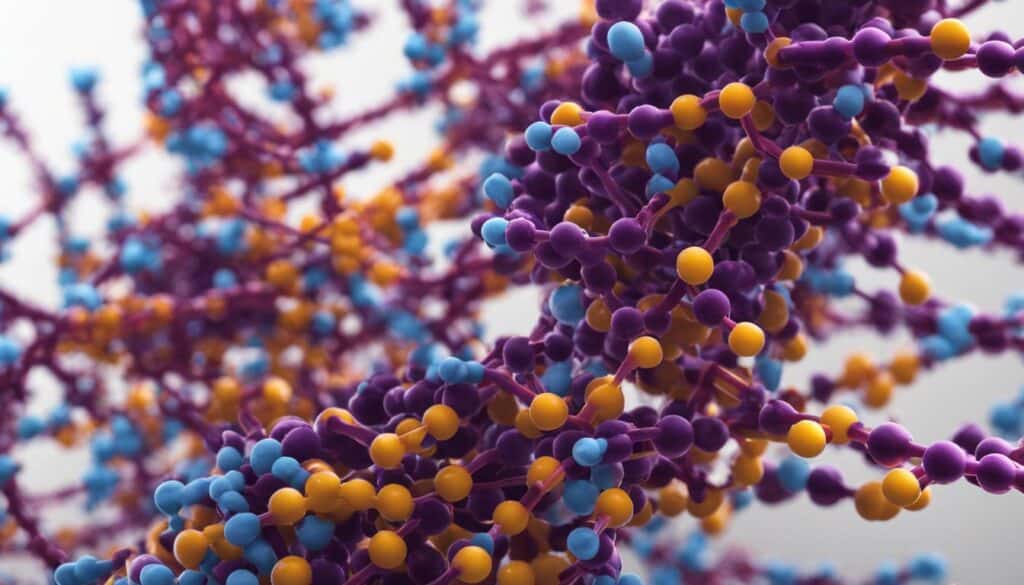
Biotinylation is a powerful technique for protein labeling, leveraging the strong binding affinity between biotin and avidin or streptavidin. This method involves the attachment of biotin molecules to proteins using biotinylation reagents. These reagents contain the biotinyl group, a spacer arm, and a reactive group that attaches to functional groups on proteins. The spacer arm plays a crucial role in determining the availability of biotin for binding to avidin or streptavidin.
Spacer arms can vary in length, affecting the accessibility of biotin for binding. Shorter spacer arms allow for stronger binding interactions, while longer spacer arms provide increased flexibility and accessibility for biotin binding.
Biotinylation offers several advantages as a labeling method. Its strong and selective binding to avidin or streptavidin proteins ensures stable and specific interactions, allowing for efficient protein detection and purification. Additionally, biotinylated proteins can be visualized using fluorophore-conjugated avidin or streptavidin, enabling imaging and localization studies.
| Advantages of Protein Labeling through Biotinylation: |
|---|
| High binding affinity between biotin and avidin/streptavidin |
| Specific and stable protein interactions |
| Efficient protein detection and purification |
| Fluorophore-conjugated avidin/streptavidin for visualization and imaging |
Real-Life Applications of Protein Labeling
Protein labeling finds extensive applications in diverse fields, including drug discovery, diagnostics, and proteomics. By attaching labels to proteins, researchers are able to track and identify specific proteins of interest, enabling a deeper understanding of their function and interactions within biological systems.
In drug discovery, protein labeling plays a critical role in the development and evaluation of potential therapeutic molecules. By labeling proteins involved in disease pathways, researchers can assess the efficacy and specificity of drug candidates, leading to the identification of promising compounds for further development.
Diagnostics also benefit greatly from protein labeling techniques. Through the use of labeled antibodies, diagnostic tests can accurately detect the presence of specific proteins associated with various diseases and conditions. This enables early detection, accurate diagnosis, and monitoring of disease progression, ultimately improving patient outcomes.
Proteomics, the study of proteins on a global scale, relies heavily on protein labeling to unravel the complex network of protein interactions within cells. By labeling proteins with fluorophores or other markers, researchers can visualize and track protein localization, movement, and interactions in real-time, providing valuable insights into cellular processes and signaling pathways.
| Application | Example |
|---|---|
| Drug Discovery | Labeling proteins involved in disease pathways to evaluate drug candidates. |
| Diagnostics | Labeled antibodies for accurate detection of disease-related proteins. |
| Proteomics | Visualizing and tracking protein interactions and localization. |
Protein labeling is a dynamic and versatile tool that continues to advance biological research and drive innovation in various fields. With ongoing advancements in labeling techniques and the development of new reagents and protocols, our understanding of proteins and their roles in health and disease will undoubtedly continue to expand.

Protein Labeling for Imaging: Unlocking the Microscopic World of Biological Processes
Protein labeling for imaging offers valuable insights into intricate biological processes and interactions at the microscopic level. Through this technique, we can visualize and study proteins within living cells, enabling a deeper understanding of their functions and behaviors. By attaching fluorescent labels to proteins of interest, researchers can track their movements, interactions, and localization with high precision.
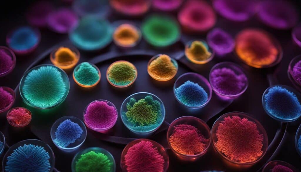
Fluorescently labeled proteins are commonly used in techniques like fluorescence microscopy, confocal microscopy, and live-cell imaging. With the help of these advanced imaging tools, we can observe dynamic processes such as protein trafficking, cell signaling, and protein-protein interactions in real-time. The ability to visualize these processes provides crucial insights into cellular function and paves the way for breakthroughs in various fields, including cell biology, neuroscience, and drug discovery.
When performing protein labeling for imaging, it is important to choose the right fluorescent label based on the experimental requirements. Different fluorophores offer varying levels of brightness, photostability, and compatibility with specific imaging systems. It is also essential to optimize labeling protocols to ensure minimal cell toxicity and maximum labeling efficiency.
The Benefits of Protein Labeling for Imaging
- Visualization of protein localization and dynamics within cells
- Study of protein-protein interactions
- Investigation of signaling pathways and cellular processes
- Validation of protein expression and targeting
- Identification of subcellular structures and organelles
As technology continues to evolve, protein labeling for imaging is becoming increasingly powerful and versatile. It allows us to delve deeper into the complex machinery of living cells, uncovering the secrets of life at a microscopic level.
| Advantages of Protein Labeling for Imaging | Challenges of Protein Labeling for Imaging |
|---|---|
| Provides real-time, dynamic information | Optimizing labeling protocols for different proteins |
| Enables visualization of subcellular structures | Selecting the most suitable fluorescent label |
| Allows for precise localization of proteins | Minimizing cell toxicity associated with labeling |
Protein labeling for imaging is revolutionizing our understanding of biology, allowing us to visualize the invisible and decipher the intricate dance of proteins within living cells. By harnessing the power of fluorescence, we can unlock the secrets of life at its most fundamental level.
Conclusion
Protein labeling techniques are indispensable tools that enable scientists to study, detect, and unravel the intricate world of proteins. In biological research, different molecules such as biotin, fluorophores, and radioactive isotopes are covalently attached to proteins to facilitate their identification and study. Biotin, in particular, is commonly used due to its strong binding to avidin and streptavidin proteins. Chemical methods, utilizing biotinylation reagents, are widely employed to attach biotin molecules to proteins. These reagents consist of the biotinyl group, a spacer arm, and a reactive group that binds to functional groups on proteins.
The length of the spacer arm plays a crucial role in determining the availability of biotin for binding to avidin or streptavidin. Enzyme labels like horseradish peroxidase and alkaline phosphatase are also frequently utilized in protein labeling. In order to generate a detectable signal, these enzymes require the addition of a substrate. Additionally, active site probes serve as specific labeling reagents designed to bind to particular enzyme active sites. These probes are instrumental in enzyme inhibition studies and activity-based proteomic profiling.
Overall, protein labeling techniques provide researchers with invaluable insights into protein function and interactions. They play a vital role in various fields, including drug discovery, diagnostics, and proteomics. Through protein labeling, scientists are able to visualize and study biological processes and interactions at a microscopic level. By unlocking the science of labeling protein, researchers can uncover the secrets of the intricate world of proteins and advance our understanding of the fundamental building blocks of life.
FAQ
Q: What is protein labeling?
A: Protein labeling is a technique used in biological research to detect and purify proteins of interest. It involves attaching different molecules, such as biotin, fluorophores, or radioactive isotopes, to proteins, allowing for their identification and study.
Q: How are proteins labeled?
A: Proteins can be labeled using various methods. Commonly, biotinylation reagents are used, which contain the biotinyl group, a spacer arm, and a reactive group that attaches to functional groups on proteins. Other methods involve enzyme labels that require the addition of a substrate to generate a detectable signal.
Q: Why is biotin a commonly used label?
A: Biotin is commonly used as a protein label because it has a strong binding affinity to avidin and streptavidin proteins. This allows for efficient detection and purification of biotinylated proteins.
Q: What are the different protein labeling techniques?
A: Protein labeling techniques include covalent attachment of molecules like biotin, fluorophores, or radioactive isotopes, as well as the use of enzyme labels. These techniques enable the visualization and study of proteins in biological systems.
Q: How do protein labeling reagents work?
A: Protein labeling reagents, such as biotinylation reagents, contain a biotinyl group, a spacer arm, and a reactive group. The reactive group attaches to functional groups on proteins, allowing for the covalent attachment of biotin molecules.
Q: What are protein labeling assays used for?
A: Protein labeling assays are used to detect labeled proteins. Enzyme labels, such as horseradish peroxidase and alkaline phosphatase, require the addition of a substrate to generate a detectable signal.
Q: How is protein labeling used in fluorescence applications?
A: Protein labeling is used in fluorescence applications by attaching fluorescent labels to proteins. This enables the visualization and imaging of proteins in biological systems.
Q: Can you explain protein labeling through biotinylation?
A: Protein labeling through biotinylation involves attaching biotin molecules to proteins using biotinylation reagents. Biotin has a strong binding affinity to avidin and streptavidin proteins, allowing for efficient detection and purification of biotinylated proteins.
Q: What are the real-life applications of protein labeling?
A: Protein labeling techniques have various real-life applications, including drug discovery, diagnostics, and proteomics. These techniques allow researchers to study protein function and interactions in different fields of research.
Q: How is protein labeling used for imaging purposes?
A: Protein labeling is used for imaging purposes to visualize and study biological processes at a microscopic level. Labeled proteins can be tracked and observed in real-time, enabling researchers to gain insights into complex biological phenomena.
How Many Carbs Are in a Protein-Packed Breakfast Burrito?
If you’re counting breakfast burrito carbs, it’s essential to be aware of your protein-packed morning meal’s carb content. The number of carbs in a protein-packed breakfast burrito can vary depending on its ingredients. While the tortilla itself contributes to the carb count, fillings like beans, eggs, and veggies may add additional carbohydrates. Make sure to check the nutrition facts or consult a dietitian for accurate carb counting.



Leave a Reply